Showing posts with label hydroxyurea. Show all posts

Tuesday, April 7, 2015

bio125, hydroxyurea induced G1 arrest

X Email ppt to students

Section 1
bio125, hydroxyurea induced G1 arrest

check FOA plate
take pictures

http://en.wikipedia.org/wiki/Hydroxycarbamide

precision medicine youtube channel
whitehouse precicion medicine
https://www.youtube.com/watch?v=RIzbg8REzGw

http://www.pha.jhu.edu/~ghzheng/old/webct/note7_3.files/F13-22b.gif


MSH2 DNA mismatch-repair mechanism

at 10am, check +HU and -HU cells under micro scrope. The key difference is the missing of S-phased budded cell in +HU treatment. Yes, HU is working.


Section 2;

Same drill, and the experiment went more smooth.

FOA results in section 2 are mostly consistent with our expectaction.


Problems: 
Some student closed off the diagpham for microscope, some did not pull the switch for the vertical adaptor. Some did not know how to focus.


Wednesday, March 18, 2015

Hydroxyurea treatment of yeast cells with WT or deletion of MSH2 gene.

Materials
  • 2M Hydroxy Urea (HU) stock solution in water: 5mL x 2M x FW 76.06/1000 = 0.7606 gram. 

HU are salt-like, and can dissolve in water in about 10 minutes at 30C with shaking. 


  • 1 ug/ul Propidum Iodine(PrI) stock
  • AGY124, yeast strain with pRS413 and pSH44 (This is the plasmid control)
  • AGY125, yeast strain with the wild type pMSH2 and pSH44 (This is the wildtype MSH2 control)


These strains were shipped from Gammie lab in January 2015,
see http://hongqinlab.blogspot.com/2015/01/bio125-strains-from-gammie-lab-media.html
Grown in SD-HIS-TRP-URA media by Dr. Kioko on Monday March 16, 2015

  • Microscope and smart-phone stages. Students can take pictures of cell morphology. 


Experimental procedure and results. 

Under microscope, WT and Vector control yeast cells are about 50% in budding phases (big cells with small buds). 

Vector control cells, about 50% in budding phases. 

WT in about 50% budding phases.

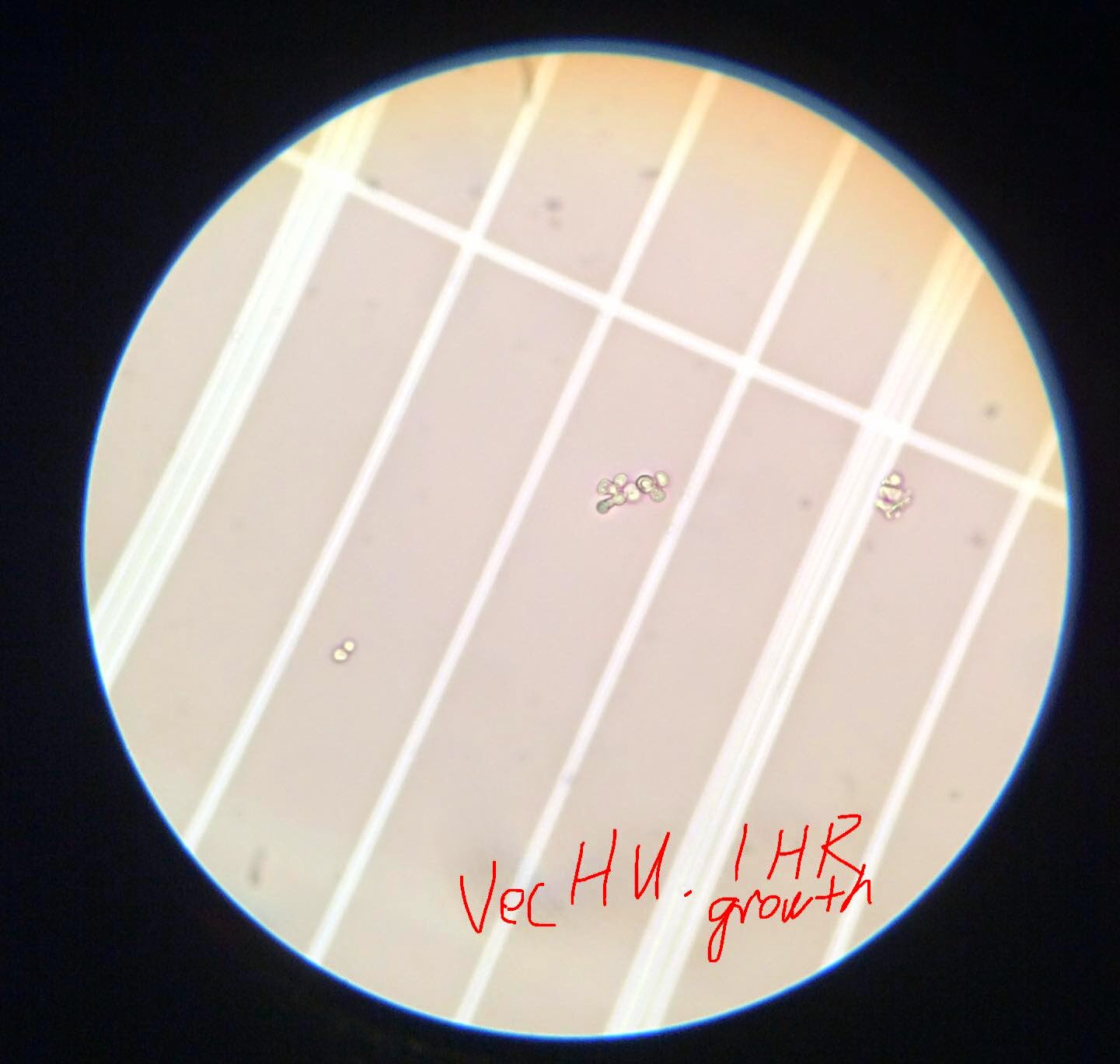
WT and Vec cells were treated with 200mM HU for 1 hour. Under microscope, it can be seen that HU-treated cells are most round and arrested at G1 phase. (Big balls in a string). 


For students:
Day1:
450ul log-culture +/- 50ul HU, monitor shape change under microscope.
After 1.5 hours, HU-arrested cells should be like watermelons.
Then wash with water once, resuspend with 500ul water, left at 4C.
(20160405. Water arrested cells just like HU, so cells ). 

Day 2.
spin down cells, add 500ul SD growth media,
shake at 30C
after 1.5 hours, monitor shape changes under microscope.
spin down, add 500ul 70% enthanol, shake at room temp

Day 2.5 Faculty do PI stain and flow cytomer run

Day 3. R exercise on flow data


References:
http://hongqinlab.blogspot.com/2013/12/sce-cell-cycle-and-morphology.html
http://hongqinlab.blogspot.com/2015/01/cell-cycle-and-msh2-project.html


Monday, March 9, 2015

yeast cell cycle flow cytometry notes


=> Change, Boone, PNAS 2002, genome-wide screen for MMS sensitive mutants
http://www.pnas.org/content/99/26/16934.full.pdf+html?with-ds=yes
MMS 0.035% vol/vol in YPD plates
HU in 0.2M in YPD
Flow cytometry: cells fixed in 70% ethanol, resuspend in 0.5ml of 0.1 mg/ml RNase A in 50mM sodium citrate, incubated at 30C O/N.  Cell were stained with 2 uM SYTOX gree (Molecular Probes) in 50mM citrate.


Thursday, January 8, 2015

Cell cycle, MSH2, mutagen treatment

A Gammie: 
"We have never observed a growth defect with the null or any of the mutants, but we think there might be some differences in the presence of DNA damaging agents (HU or cisplatin) – you could try that for your flow experiments. I wonder if your strains have picked up secondary mutations. We never propagate the strains for very long because they are mutators and pick up deleterious mutations during propagation."

Gammie used HU and MMS in her 2013 DNA repair paper
http://www.sciencedirect.com/science/article/pii/S1568786412002819
HU was 0.1M = 100mM and MMS was 0.04% in this paper. Treatments were 1.5 hours (about one generation time for the BY lab strain).

Hydroxyurea (HU) (no respiratory risk label on Sigma)
AKA, hydroxycarbamide
http://en.wikipedia.org/wiki/Hydroxycarbamide  
http://www.sigmaaldrich.com/catalog/product/sigma/h8627?lang=en&region=US
http://www.sigmaaldrich.com/content/dam/sigma-aldrich/docs/Sigma/Product_Information_Sheet/2/h8627pis.pdf
FW: 76.06, 
So, 1 gram can make  2M solution of 1000x2 /76.06 mL = 13.1/2 = 6.5 mL  (This is 1000/6.5 mg/mL = 154 mg/mL)


"Hydroxyurea is freely soluble in water, to at least 50 mg/ml (5%). However, because hydroxyurea decomposes in the presence of moisture, aqueous solutions are probably not stable. It is recommended to store hydroxy-urea at 2-8 °C. Hydroxyurea should be stored in a dry atmosphere in airtight containers." (Sigma) 


See cell cycle arrest protocol form https://sites.google.com/site/mckeogh2/protocols

Wikipedia entry shows that hydroxurea is a WHO essential medicine, and used in some leukemia treatment.


Hydroxyurea (Hydroxycarbamide) ab142613 - Abcam

www.abcam.com/Hydroxyurea-Hydroxycarbamide-ab142613.pdf
Claim 100mM stock solution in water or DMSO? 


http://www.genetics.org/content/189/2/533.full.pdf+html
Tripathi2011Genetics:  HU cause G2/M arrest in yeast cells.


====================

Methyl methanesulfonate (MMS) (respiratory risk, require facial mask)

Molar mass110.13 g/mol
So, 1 gram make 1000x1 /110.13 mL = 9.08 mL
http://www.sigmaaldrich.com/catalog/product/aldrich/129925?lang=en&region=US
http://en.wikipedia.org/wiki/Methyl_methanesulfonate


Cisplatin (very expensive at Sigma)

Need to know to their dosage to conclude which one is actually more expensive to do experiments. 

References:
http://hongqinlab.blogspot.com/2013/12/sce-cell-cycle-and-morphology.html