Kioko: 1:100 in 3 days
Mon 4pm, 1:30 dilution for 8am section.
Showing posts with label MSH2. Show all posts
Showing posts with label MSH2. Show all posts
Monday, April 6, 2015
Thursday, March 26, 2015
*** Calibur, gating for wildtype and vector for bio125 MSH2 yeast strains
__ AGY 124 (vector control), AGY125(MSH2 control), grown in SD-His-Trp-Ura for 2 days
__ take 100ul to 2ml water
__ point sonication, level 2, 4 times
Calibur, E03 FSC, 952 SSC, choose gate R1 for yeast cells.
template file: tempalte20150320-R1R2-v2

__ take 100ul to 2ml water
__ point sonication, level 2, 4 times
Calibur, E03 FSC, 952 SSC, choose gate R1 for yeast cells.
template file: tempalte20150320-R1R2-v2

Wednesday, March 18, 2015
Hydroxyurea treatment of yeast cells with WT or deletion of MSH2 gene.
Materials
- 2M Hydroxy Urea (HU) stock solution in water: 5mL x 2M x FW 76.06/1000 = 0.7606 gram.
 |
| HU are salt-like, and can dissolve in water in about 10 minutes at 30C with shaking. |

- 1 ug/ul Propidum Iodine(PrI) stock
- AGY124, yeast strain with pRS413 and pSH44 (This is the plasmid control)
- AGY125, yeast strain with the wild type pMSH2 and pSH44 (This is the wildtype MSH2 control)
 |
| These strains were shipped from Gammie lab in January 2015, see http://hongqinlab.blogspot.com/2015/01/bio125-strains-from-gammie-lab-media.html |
 |
| Grown in SD-HIS-TRP-URA media by Dr. Kioko on Monday March 16, 2015 |
- Microscope and smart-phone stages. Students can take pictures of cell morphology.
Experimental procedure and results.
Under microscope, WT and Vector control yeast cells are about 50% in budding phases (big cells with small buds).
 |
| Vector control cells, about 50% in budding phases. |
 |
| WT in about 50% budding phases. |
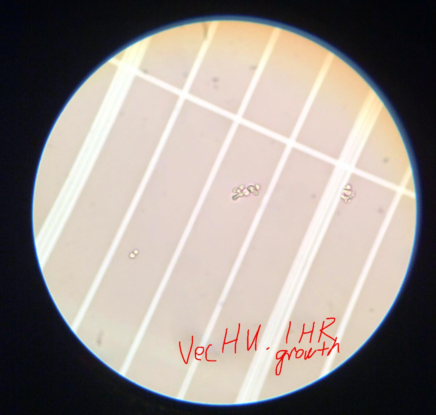
WT and Vec cells were treated with 200mM HU for 1 hour. Under microscope, it can be seen that HU-treated cells are most round and arrested at G1 phase. (Big balls in a string).


For students:
Day1:
450ul log-culture +/- 50ul HU, monitor shape change under microscope.
After 1.5 hours, HU-arrested cells should be like watermelons.
Then wash with water once, resuspend with 500ul water, l
(20160405. Water arrested cells just like HU, so cells ).
Day 2.
spin down cells, add 500ul SD growth media,
shake at 30C
after 1.5 hours, monitor shape changes under microscope.
spin down, add 500ul 70% enthanol, shake at room temp
Day 2.5 Faculty do PI stain and flow cytomer run
Day 3. R exercise on flow data
References:
http://hongqinlab.blogspot.com/2013/12/sce-cell-cycle-and-morphology.html
http://hongqinlab.blogspot.com/2015/01/cell-cycle-and-msh2-project.html
Tuesday, January 13, 2015
Bio125, strains from Gammie lab
Received on Jan 13, 2015
AGY125, yeast strain with the wild type pMSH2 and pSH44 (This is the wildtype MSH2 control)
Yeast Media:
SC-His-Trp
-HIS to keep pMSH2
-TRP to keep pSH44 reporter plasmid
Ecoli Media:
LB + amp because pRS413 has a beta-lactamase which offer resistance to beta-lactam antibiotics such as ampicillin and penicillin.
I added 5ul of 50 mg/mL ampicilin stock to 5ml LB.
Amplicilin, histidine stocks, LB and SC-His-Trp media are provided by Dr. Kioko.
20150113, 4pm, yeast liquid sample were at 30C shaker, and Ecoli strains at 37C shaker.
The yeast plate of AGY124 and 125 were at 30C incubator.
Plate AGY75 were left outside for histidine to dry.
20150114. 10am. Liquid growth are obvious for both yeast and Ecoli strains. Kioko froze the Ecoli in 20% glycerol as stocks.


Observation:
AGY124 and AGY125 grew on SD-Trp-His-Ura plates. They form pink colonies when speaded dense population.
2015 Feb 12, I finally obtained SD-Trp-Ura plates, and spread AGY75 strains from the glass-tube cultures.
2015 Feb 16. AGY75 grows well on SD-Trp-Ura plate (Problem: some colonies became pink, but some remain white after a while. ).

2015 Feb 27 Fri, 9pm. Streaks AGY75 from Box10 G3 to SD-URA-TRP+HIS plate. Left at 30C incubator in Sci 246.

Yeast strains
AGY 75, yeast strain with pSH44 reporter plasmid
AGY124, yeast strain with pRS413 and pSH44 (This is the plasmid control)
Ecoli strain with plasmid
AG372, pmsh2-H658R
AG421, pmsh2-A618V
Yeast Media:
To grow AGY125 and AGY124
-HIS to keep pMSH2
-TRP to keep pSH44 reporter plasmid
To grow AGY75, SC-Trp
I added 2.5ul 88mg/ml histidine stock to 5ml SC-His-Trp for a final concentration of 44ug/ml based on Gammie protocol according to Dr. Kioko
For plate growth, I dripped 1X histitine to the SC-HIS-Trp plate for AGY75
Ecoli Media:
LB + amp because pRS413 has a beta-lactamase which offer resistance to beta-lactam antibiotics such as ampicillin and penicillin.
I added 5ul of 50 mg/mL ampicilin stock to 5ml LB.
Amplicilin, histidine stocks, LB and SC-His-Trp media are provided by Dr. Kioko.
20150113, 4pm, yeast liquid sample were at 30C shaker, and Ecoli strains at 37C shaker.
The yeast plate of AGY124 and 125 were at 30C incubator.
Plate AGY75 were left outside for histidine to dry.
20150114. 10am. Liquid growth are obvious for both yeast and Ecoli strains. Kioko froze the Ecoli in 20% glycerol as stocks.
Observation:
AGY124 and AGY125 grew on SD-Trp-His-Ura plates. They form pink colonies when speaded dense population.
2015 Feb 12, I finally obtained SD-Trp-Ura plates, and spread AGY75 strains from the glass-tube cultures.
2015 Feb 16. AGY75 grows well on SD-Trp-Ura plate (Problem: some colonies became pink, but some remain white after a while. ).
2015 Feb 27 Fri, 9pm. Streaks AGY75 from Box10 G3 to SD-URA-TRP+HIS plate. Left at 30C incubator in Sci 246.
 |
| Same plate, front view |
Reference:
http://hongqinlab.blogspot.com/2013/01/yeast-msh2-project-in-undergraduate.html
http://hongqinlab.blogspot.com/2015/02/bio125-yeast-transformation.html
http://hongqinlab.blogspot.com/2015/02/bio125-yeast-transformation.html
Thursday, January 8, 2015
Cell cycle, MSH2, mutagen treatment
A Gammie:
"We have never observed a growth defect with the null or any of the mutants, but we think there might be some differences in the presence of DNA damaging agents (HU or cisplatin) – you could try that for your flow experiments. I wonder if your strains have picked up secondary mutations. We never propagate the strains for very long because they are mutators and pick up deleterious mutations during propagation."
Gammie used HU and MMS in her 2013 DNA repair paper
http://www.sciencedirect.com/science/article/pii/S1568786412002819
HU was 0.1M = 100mM and MMS was 0.04% in this paper. Treatments were 1.5 hours (about one generation time for the BY lab strain).
Hydroxyurea (HU) (no respiratory risk label on Sigma)
AKA, hydroxycarbamidehttp://en.wikipedia.org/wiki/Hydroxycarbamide
http://www.sigmaaldrich.com/content/dam/sigma-aldrich/docs/Sigma/Product_Information_Sheet/2/h8627pis.pdf
FW: 76.06,
So, 1 gram can make 2M solution of 1000x2 /76.06 mL = 13.1/2 = 6.5 mL (This is 1000/6.5 mg/mL = 154 mg/mL)
"Hydroxyurea is freely soluble in water, to at least 50 mg/ml (5%). However, because hydroxyurea decomposes in the presence of moisture, aqueous solutions are probably not stable. It is recommended to store hydroxy-urea at 2-8 °C. Hydroxyurea should be stored in a dry atmosphere in airtight containers." (Sigma)

Hydroxyurea (Hydroxycarbamide) ab142613 - Abcam
www.abcam.com/Hydroxyurea-Hydroxycarbamide-ab142613.pdf
Claim 100mM stock solution in water or DMSO?
http://www.genetics.org/content/189/2/533.full.pdf+html
Tripathi2011Genetics: HU cause G2/M arrest in yeast cells.


====================
Methyl methanesulfonate (MMS) (respiratory risk, require facial mask)
| Molar mass | 110.13 g/mol |
http://www.sigmaaldrich.com/catalog/product/aldrich/129925?lang=en®ion=US
http://en.wikipedia.org/wiki/Methyl_methanesulfonate
Cisplatin (very expensive at Sigma)
Need to know to their dosage to conclude which one is actually more expensive to do experiments.
References:
http://hongqinlab.blogspot.com/2013/12/sce-cell-cycle-and-morphology.htmlThursday, April 17, 2014
Design a mutagenic primer to engineer human C333F mutation to yeast MSH2
Goal: Engineer the human C333F mutation into yeast MSH2.
Link of video tutorial of this exercise: https://youtu.be/SWgE_PfxmeU
Video on G548C is https://youtu.be/6C8E_OZKeeQ
The principle of site specific mutagenesis can be found in this tutorial video. Explanation of cognant sites can be see in this Youtube video.
Copy yeast and human MSH2 protein sequences in FASTA format from this link.

Google "EMBL ClustalW2". Then, paste the yeast and human MSH2 sequences in FASTA format into ClustalW2 window.

After "submit", you should see the protein alignment.

Identify the cognant site in yeast MSH2. Human C333 corresponds to yeast C345.

Therefore, human mutation C333F should become C345F mutation in yeast MSH2 gene.
Now, we can design the mutagenic primer based on the DNA coding sequences of the yeast MSH2 (linked here). You can copy-paste this sequences into ApE.
In the yeast MSH2 ORF sequence, the 345th amino acid position corresponds to 345x3=1035 nucleotide position. So, the codon position is 1033-1035. We can select these 3 nucleotide in ApE to and double check this codon.

This selected codon should be:
 We can verify that codon TGC is amino acid "C".
To design a mutagenic primer, we will pick 10 bp left to the TGC codon and 10 bp right to the TGC codon, by select from 1023-1045.


Copy the selected sequence into a new ApE window.

At the center of this sequence should be "TGC".

The yeast codon usage table is:

The preferred codon for "F" in yeast is "TTT".
We can manually change "TGC" to "TTT".

We can save this file in ApE as "C345F_mutagenic_primer.seq".

Link of video tutorial of this exercise: https://youtu.be/SWgE_PfxmeU
Video on G548C is https://youtu.be/6C8E_OZKeeQ
The principle of site specific mutagenesis can be found in this tutorial video. Explanation of cognant sites can be see in this Youtube video.
Copy yeast and human MSH2 protein sequences in FASTA format from this link.
Google "EMBL ClustalW2". Then, paste the yeast and human MSH2 sequences in FASTA format into ClustalW2 window.
After "submit", you should see the protein alignment.
Identify the cognant site in yeast MSH2. Human C333 corresponds to yeast C345.
Therefore, human mutation C333F should become C345F mutation in yeast MSH2 gene.
Now, we can design the mutagenic primer based on the DNA coding sequences of the yeast MSH2 (linked here). You can copy-paste this sequences into ApE.
In the yeast MSH2 ORF sequence, the 345th amino acid position corresponds to 345x3=1035 nucleotide position. So, the codon position is 1033-1035. We can select these 3 nucleotide in ApE to and double check this codon.
This selected codon should be:
To design a mutagenic primer, we will pick 10 bp left to the TGC codon and 10 bp right to the TGC codon, by select from 1023-1045.
Copy the selected sequence into a new ApE window.
At the center of this sequence should be "TGC".
The yeast codon usage table is:
The preferred codon for "F" in yeast is "TTT".
We can manually change "TGC" to "TTT".
We can save this file in ApE as "C345F_mutagenic_primer.seq".
Thursday, April 10, 2014
BIO125, research day poster on H658Y
FOA plate show that H658Y has as few colonies as potential wildtype MSH2 control. So, H658Y has nearly-normal MMR function. The mutation occurs in ATPase domain, so maybe human MSH2-H658Y just cannot repair as efficiently as the wildtype one, even though its actual repair machinery is normal.
It is also possible that protein expression level of H658Y is lowered.
In Gammie 2007, H658Y was shown to be '-' on MMR. So, our FOA results are not consistent with Gammie07. It is possible that H658Y cells were substantially lower than wildtype MSH2.
It is also possible that protein expression level of H658Y is lowered.
In Gammie 2007, H658Y was shown to be '-' on MMR. So, our FOA results are not consistent with Gammie07. It is possible that H658Y cells were substantially lower than wildtype MSH2.
Sunday, March 9, 2014
yeast MSH2 gene expression peaks in G1 phase (protein peak in S phase?)
MSH2 Expression and yeast cell cycle
The yeast MSH2 mRNA peak in G1 phase. If the MSH2 protein have half-live longer than G1 phase, the protein MSH2 level can be high in both G1 and S phases.
From:
http://genome-www.stanford.edu/cgi-bin/cellcycle/drawCCquery.pl
The original paper, Cho 1998


cell cycle and gene expression:
http://www.nature.com/nrm/journal/v2/n11/box/nrm1101-815a_BX1.html

At GEO, GSE3635, two cell cycle data of wildtype W303
The GEO Dataset Browser allow user to look at gene name and its expression profile. I did not find a way to generate a heatmap?!
The yeast MSH2 mRNA peak in G1 phase. If the MSH2 protein have half-live longer than G1 phase, the protein MSH2 level can be high in both G1 and S phases.
http://genome-www.stanford.edu/cgi-bin/cellcycle/drawCCquery.pl
The original paper, Cho 1998
cell cycle and gene expression:
http://www.nature.com/nrm/journal/v2/n11/box/nrm1101-815a_BX1.html
At GEO, GSE3635, two cell cycle data of wildtype W303
The GEO Dataset Browser allow user to look at gene name and its expression profile. I did not find a way to generate a heatmap?!
Saturday, March 8, 2014
human MSH2 using the UCSC genome browser
Go to UCSC genome browser: http://genome.ucsc.edu/
Input "MSH2" to the "search term" and press "submit".

You will see 6 entries of human MSH2. You can tell which one is the longest form based on their coordinates. Click any one of them will take you to the genomic regions of hMSH2


For 'Phenotype and Literature', we can 'hide' Publications and show'full' 'pack'ed ClinVar Variants.

After hitting 'refresh', we can see many 'mutations' in the human MSH2 region.

Go to 'Tools' on the menu toolbar, and select 'Table Browser'

Make sure that we are still at the MSH2 genomic region, chr2:47630206-47710367.
We can set 'group' as 'Phenotype and Literature', 'track' as "clinVar Variants', and 'table' as "ClinVar Main(clivarmain)', The 'region' should select 'position' in chr2:47630206-47710367,
the input output file as "human_msh2_clinvariant". Then click "get output".


Human MSH2 protein: NP_000242
http://genome.ucsc.edu/training.html
Click "genomes"

You will see 6 entries of human MSH2. You can tell which one is the longest form based on their coordinates. Click any one of them will take you to the genomic regions of hMSH2
The region of MSH2 in the human genome looks like:
Scroll down, and we can see options for adjustment.
For "Genes and Gene Predictions", we can 'hide' UCSC genes and 'pack' RefSeq Genes'. Hit 'refresh' and we should see the updated genome view.
For 'Phenotype and Literature', we can 'hide' Publications and show
After hitting 'refresh', we can see many 'mutations' in the human MSH2 region.
Go to 'Tools' on the menu toolbar, and select 'Table Browser'
Make sure that we are still at the MSH2 genomic region, chr2:47630206-47710367.
We can set 'group' as 'Phenotype and Literature', 'track' as "clinVar Variants', and 'table' as "ClinVar Main(clivarmain)', The 'region' should select 'position' in chr2:47630206-47710367,
the input output file as "human_msh2_clinvariant". Then click "get output".
The downloaded file is a text file, and we can open it with any text editor.
At the stage, we can run Excel and generate a new spread sheet. We can then copy-paste the downloaded clinic variants of human MSH2 into the Excel sheet.
The file now looks like
In the column 'clinSign', we can see the clinic relevance of the mutations. Let pick a 'pathogenic' variant start at 47702268. From column 'hgvsProt', we can see this is a mutation Pro622Leu (or P622L).
To double-check the SNP rs2303426, we can look at NCBI db-SNP database:
Now, we can design a mutagenic primer that can introduce this human mutation P622L into the cognant site in yeast. The mutagenic primer should also consider the preferred codons in yeast.
We shall also be able to design PCR primers that can amplify a fragment that cover this mutation in yeast MSH2 and identify a Restriction Enzyme that can distinguish the wildtype yeast MSH2 from the mutant MSH2.
Yeast codon usage can be found at:
http://www.genscript.com/cgi-bin/tools/codon_freq_table
References:
http://genome.ucsc.edu/cgi-bin/hgTracks?db=hg19&position=chr2%3A47636726-47690166Human MSH2 protein: NP_000242
http://www.genscript.com/cgi-bin/tools/codon_freq_table
http://genome.ucsc.edu/training.html
Subscribe to:
Posts (Atom)